A single injection of our Ebola vaccine provided 100% protection for mice exposed to Ebola virus.
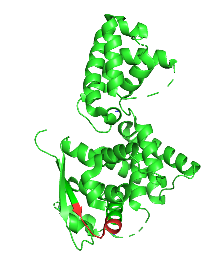
The vaccine creates T-Cells that attack NP44-52, a specific segment of the Ebola nucleocapsid protein, identified by the red arrow in the picture.
For practical purposes, Marburg is best thought of as a weaponized version of Ebola.
By relying on T-cells rather than antibodies, FlowVax Ebola/Marburg provides long lasting protection through enhanced immune targeting and surveillance of the hidden Ebola and Marburg virus nucleocapsid proteins. This protection can last multiple years.
FlowVax Marburg Vaccine Development has been funded by the United States Department of Defense Chemical and Biological Defense Program.
Key Features
- Under Development, FDA Approval Required.
- USA National Laboratory Study shows 100% Efficacy in C57BL/6 Mice.
- Research Paper: An effective CTL peptide vaccine for Ebola Zaire Based on Survivors’ CD8+ targeting of a particular nucleocapsid protein epitope with potential implications for COVID-19 vaccine design. [PDF] [Publisher Website].
- Non-human primate study for Marburg protection was funded by United States Department of Defense.
Current Status
-
PRE-CLINICAL FEASIBILITY
-
PRE-PHASE I CLINICAL STUDY
-
PHASE I
What is Ebola?
Ebola, also known as Ebola virus disease (EVD) or Ebola hemorrhagic fever (EHF), is a viral hemorrhagic fever of humans and other primates caused by ebolaviruses.
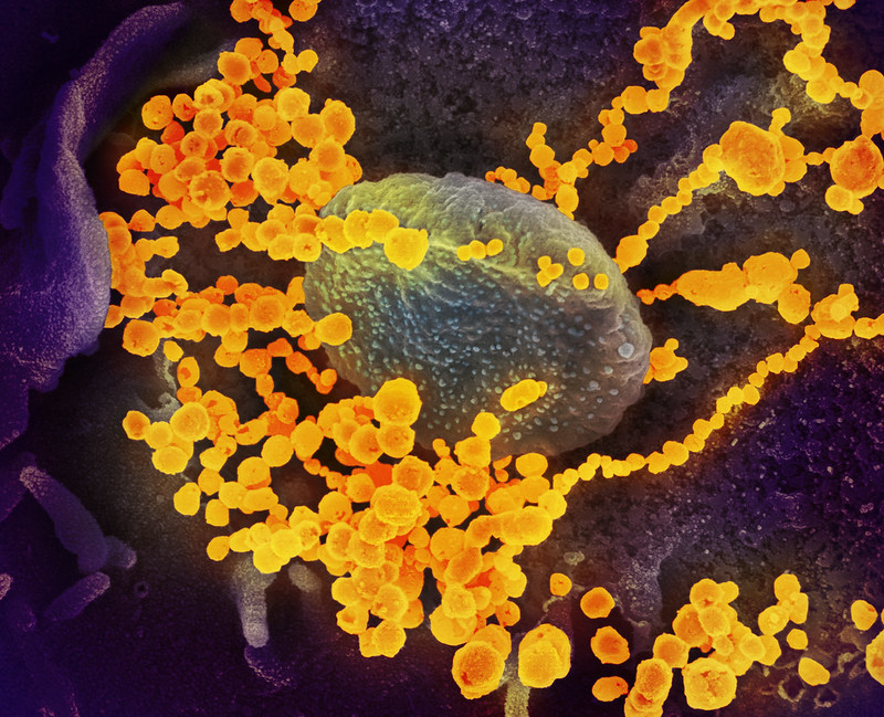
Colorized scanning electron micrograph of filamentous Ebola virus particles (green) attached to and budding from a chronically infected VERO E6 cell (blue) (25,000x magnification). Image captured and color-enhanced at the NIAID Integrated Research Facility in Ft. Detrick, Maryland. Credit: NIAID
What is Marburg?
Marburg virus is a a Category A Bioterrorism Agent according to the Centers for Disease Control. Marburg virus (MARV) causes Marburg virus disease in humans and nonhuman primates, a form of viral hemorrhagic fever. The virus is considered to be extremely dangerous.
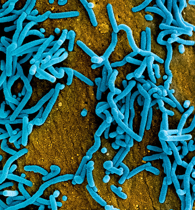
Colorized scanning electron micrograph of Marburg virus particles (blue) both budding and attached to the surface of infected VERO E6 cells (orange). Image captured and color-enhanced at the NIAID Integrated Research Facility in Fort Detrick, Maryland. Credit: NIAID


